Posted by: Metrolina Eye Associates in Diabetic Retinopathy
Most people are aware of the growing number of Americans with diabetes. However, most are not aware of the serious affects diabetes can have on the eyes. In fact, according to the CDC, the leading cause of blindness for Americans age 20-74 is something called diabetic retinopathy.
Diabetic retinopathy is an eye disease that can be caused by diabetes. It is characterized mainly by progressive damage to the blood vessels within the retina (the back inside layer of the eye). Proper functioning of the retina is critical for good clear vision. As these retinal vessels are damaged they may begin to leak blood and fluid. This is a condition called Non-Proliferative Diabetic Retinopathy, or NPDR.
There are three stages of NPDR; mild, moderate, and severe. The mild stage consists of only subtle changes to the retinal vessels and there may not be any noticeable changes to one’s vision. This is, however, a critical stage for discovering retinopathy before it progresses to the moderate and severe stages. As progression occurs more vessels throughout the retina are affected causing blood and fluid leakage to become more widespread. This can then lead to swelling between the layers of the retina, especially in the central area known as the macula. Swelling in the macula is called Diabetic Macular Edema, and is a major cause of vision loss from diabetes.
If NPDR is not properly diagnosed and managed, it may then progress to something called Proliferative Diabetic Retinopathy, or PDR. PDR is by far the most severe level of diabetic retinopathy and has devastating visual consequences. In PDR the damage to retinal vessels causes a situation where the retina is not getting the nutrients it needs. To combat this, new small vessels begin to grow within the retina, a process called neovascularization. These new vessels are very weak and susceptible to rupture. When rupture occurs relatively large amounts of blood can leak and possibly spread into the gel that fills the eyeball called vitreous. This is called a vitreous hemorrhage and can cause large floaters or shadows in the vision. Scar tissue or fibrosis can also occur, leading to something called a tractional retinal detachment. Depending on the detachment’s severity and location, it can potentially cause permanent blindness.
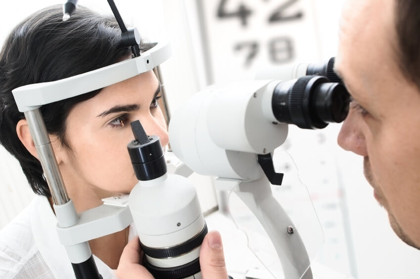
With all that can go wrong inside the eye from diabetes, it becomes abundantly clear why prevention is key and diabetic eye exams are so important. For a diabetic eye exam the parts of the eye that can possibly be affected by the disease are looked at very carefully. The eyes are always dilated and a very thorough evaluation of the retina and it’s blood vessels is performed. To aid in this process, retinal images are taken with a high definition camera. If any changes are seen then a scan of the retina called an OCT (Optical Coherence Tomography) may also be performed. This test allows visualization of the different layers of the retina to better determine the extent of any swelling or damage. Finally, a detailed report is sent to the primary care doctor or endocrinologist managing treatment. Communication of the findings with them is key because it allows them a better understanding of how well the disease is controlled and if any adjustments in medication or insulin may be required.
If during the course of a diabetic eye exam any retinopathy is found, the treatment depends on severity. In cases of NPDR without macular edema the only treatment may be close observation and strict control of blood sugar levels. If diabetic macular edema is present, then a laser procedure called focal laser may be performed. In some cases injections with a needle directly into the eye are necessary to reduce swelling before further damage occurs. In more serious cases where PDR is present treatment must be swift and aggressive. Usually a combination of extensive laser treatments and eye injections are used. With any type of diabetic retinopathy though, the most important treatment is strict control of blood glucose levels. Without that, the risk for severe and permanent vision loss remains.
It’s recommended that a diabetic eye exam is performed at least once a year, but it may be necessary every 6 months or even more often if any complications are found. If you, or someone you know, is diabetic and have not had a thorough diabetic eye exam be sure to call our office to schedule an appointment today.
By: